
Advancements in Medical Technology: A New Era in Cancer Detection and Treatment
The landscape of medical technology is rapidly evolving, particularly in the realm of cancer research and treatment. Recently, the Cancer Prevention & Research Institute of Texas (CPRIT) made headlines by awarding over $2 million in grants to two pioneering engineers from the University of Texas at Dallas. This funding is set to bolster groundbreaking research aimed at enhancing the early detection of oral cancers and overcoming one of the most significant barriers in spinal treatment: the blood-spinal cord barrier.
Innovative tools for cancer diagnosis are on the horizon.
Portable Solutions for Oral Cancer Detection
Dr. Baowei Fei, a professor of bioengineering and the Cecil H. and Ida Green Chair in Systems Biology Science at UT Dallas, has received a substantial grant of $1 million. His research focuses on creating a handheld hyperspectral imaging device capable of detecting oral and oropharyngeal cancers at their nascent stages. The device works by utilizing advanced technology that analyzes how biological cells absorb and reflect light across various wavelengths, which is beyond the human visual capabilities.
With the incorporation of artificial intelligence, this innovative tool will revolutionize the diagnostic process by identifying suspicious masses that may require further medical evaluation and treatment. Dr. Fei is collaborating with esteemed institutions, such as Texas A&M University School of Dentistry and UT Southwestern Medical Center, to ensure thorough clinical testing of the device, which is anticipated to greatly reduce the late-stage diagnoses that plague so many cancer patients today.
“The current methods of screening oral cancers are largely visual, performed under normal light, leading to late-stage discoveries that severely impact survival rates,” Dr. Fei remarked regarding the importance of this development.
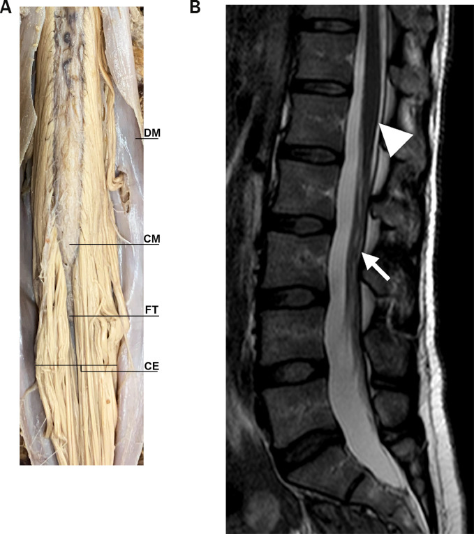
Breaking Barriers in Spinal Cord Medication Delivery
The challenges in treating spinal cord tumors stem from the complex structure known as the blood-spinal cord barrier, which functions as a protective shield but also limits medication accessibility. This barrier significantly impedes the delivery of therapeutics to spinal tissues, a problem that Dr. Zhenpeng Qin, an associate professor in mechanical engineering and bioengineering, aims to tackle. Also awarded a grant of $1 million, Dr. Qin is spearheading research to develop a novel technique that utilizes light stimulation to temporarily open this barrier, allowing effective drug delivery directly to spinal tumors.
This innovative approach is crucial as it seeks to address the limited therapy options currently available. “Our approach is unique due to its minimal invasiveness, paving the way for more effective treatments,” says Dr. Qin. His collaboration with UT Southwestern involves preclinical testing on mice, demonstrating the viability and effectiveness of this groundbreaking method.
 New methodologies are being developed for effective treatment delivery to spinal cord tumors.
Enhancing Diagnostic Imaging with Ferumoxytol
As the fields of cancer detection and treatment evolve, so too does the technology utilized in imaging. One such advancement is the use of Ferumoxytol, a superparamagnetic iron oxide nanoparticle that enhances MRI quality and offers an alternative to traditional gadolinium-based contrast agents. The approval of Ferumoxytol for clinical use since 2009 has significantly reshaped imaging practices, providing a safer profile with higher relaxivity rates and longer blood pool half-lives.
Recent discussions in the National Science Review highlight the potential of Ferumoxytol-enhanced MRI (FE-MRI) in clinical diagnostics, especially in areas such as cardiovascular disease and cancer. This technology is not only improving imaging clarity but also reducing potential risks associated with prior agents, marking a remarkable leap forward in MRI practices.
The Trajectory of Medical Imaging Advancement
The advancements surrounding FE-MRI redirect the focus towards integration and application of cutting-edge technology in clinical settings. Researchers are engaging with new themes such as micrometer-scale imaging, multimodal image fusion, and vascular segmentation, all of which underscore the interdisciplinary approach required to tackle contemporary medical challenges. As these technological innovations mature, they promise enhanced non-invasive exploration of human anatomy and more efficient diagnostics overall.
Advancements in imaging technology are paving the way for better diagnosis and treatment options.
Conclusion
The integration of new technologies into the medical field represents a transformative step forward. With innovations stemming from the University of Texas at Dallas and the utilization of superior imaging agents like Ferumoxytol, we stand on the cusp of a new era in cancer detection and treatment. As these researchers continue to push the boundaries of what is possible, their efforts not only contribute to more effective therapies but also foster hope for millions affected by these life-threatening diseases.
In summary, the advancements in early detection methodologies, breakthrough delivery techniques, and enhanced imaging technologies have the potential to reshape the cancer treatment landscape, emphasizing the criticality of ongoing investment in research and development within this vital sector.


